Tuesday, April 29, 2008

Yet another example of "non pain medicine"

We continue to see patients who have had CT guided injections for spinal pain on wrong indications and with anatomically inappropriate techniques. We will continue to publish the worst of these cases of "non pain medicine". 

This patient had neck pain on the left side irradiating to the scapular region after having been treated with a violent cervical rotation manipulation for a torticollis three years earlier. Her MRI demonstrated some insignificant disc bulging at the C3/4 level. The radiologist doing the MRI proposed a CT guided transforaminal injection of steroids which he subesequently performed as illustrated in the next two images.

The needle has been introduced from behind on the left side towards the transverse process of C3. The root hole opens laterally-downwards. An appropriate needle pass would have been 90 degrees to the actual needle via an anterior approach and going slightly cranially, sliding up along the posterior/inferior aspect of the root hole, caudal to the ganglion and posterior to the vertebral artery. The needle tip shold not pass beyond the mid-width of the articular collumn as seen in a perfect a-p projection. 
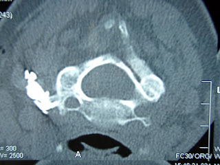
Injection of contrast fails to reach the target; there is no penetration into the spinal canal and the nerve root is not being visualized. Contrast spreads backwards along the needle through the regions of the anterior, median and posterior scalene muscles and the splenius muscle of the head. 

Nedless to say, there was no therapeutic effect of the steroids. The patient was later diagnosed in our centre with pain from the left C5/6 and C6/7 zygapophysial joints. She was treated with radiofrequency neurotomy of the medial branches responsible for the innervation of these joints and her pain has now stabilized, 6 months after treatment, at a very acceptable level of 1-2/10, down from 7-8/10 before therapy. 

Wednesday, April 23, 2008

News on our mesothelioma patient

Unfortunately, the beneficial effect on pain produced by the thoracic sympathectomy did not last longer than 3 weeks after our positive 2-week control (see our message from March 12). However, the beneficial effect on transpiration in the pain-full area remains intact, indicating that the pain that returned might be caused by other mechanisms than tumour infitration of the thoracic sympathetic chain. We therefore sent the patient for an MRI of the thoracic spine, from which the following images have been extracted.


The MRI demonstrates infiltration around the nerve root T6/7 on the left side, reaching the spinal canal without infiltrating the dural sac. There is also mesothelioma infiltration around the sympathetic chain and into the left pedicle of T6.

The patient was given a transforaminal epidural injection of a mixture of 1 cc bupivacaine 0.5% and 20 mg of depomedrone at the level of T6/7 on the left side. This test treatment completely removed his pain for 48 hours. This strongly suggests that the infiltration of tumour at this level produces the pain.

The patient is presently undergoing radiotherapy directed at the spine in an attempt to reduce the radicular pain. If this treatment is ineffective we will proceed to implant a pump system delivering bupivacaine intrathecally. If this implantation is done, the catheter tip will be placed at T5, if possible on the left side to ensure appropriate anesthesia at the spinal emergence of the T6/7 root

We will keep you informed about the evolution in subsequent messages.

Tuesday, April 22, 2008

A too long history of left sided L5 sciatica

This is a 50 y old woman with a 10 y history of intermittent low back pain, whom we saw for the first time a little over a month ago. A year ago she started to have pain irradiating along the L5 segment on the left side at the same time as her back pain became constantly present. The irradiation into the left lower extremity was described as electrical and was clearly position dependent. She also described periods when her lumbar spine became suddenly blocked, forcing her to spend a couple of days in bed till the blockade resolved. She consulted her GP who told her to loose weight and sent her for physiotherapy and osteopathy, none of which was of any benefit. After almost a year of suffering she decided to see another physician who immediately sent her for an MRI, the result of which is shown on the subsequent to images.


A large cyst emerging from the L4/5 facet joint on the left side, displacing the contents of the dural sac to the right.


On the sagittal projection on can observe the cyst and also an antelisthesis of L4 on L5 with a segmental instability L4/5, as later documented by functional images. A moderate spinal stenosis was also observed just below the L4/5 disc. The cyst and the instability in the same segment probably contributed to produce a dynamic spinal stenosis of a considerable degree.

After a thorough discussion with our spine surgeons it was decided to attempt a puncture of the facet joint cyst despite its difficult location anterior to the lamina of L5.


On this a-p film one observes the facet joint filled with contrast and to the right at its base the cyst behind the lamina. 

A needle could be inserted from a cranial direction into the cyst, from which we subsequently drained 1.5 cc of sanguinolent fluid. Twenty mg of depomedone were injected into the facet joint in an attempt to reduce a presumed inflammatory reaction likely to be responsible for the cyst formation.

The patient was seen back one month after the procedure and reported complete disappearance of her sciatica and return to her old pattern of back pain from several years back, starting a couple of days after the procedure. She was informed that there is a risk of return of symptoms since the cyst might fill up again. She was told this might require another puncture.

We have a large experience taking care of patients with facet joint cysts. These are some interesting statistics collected prospectively over the past 9 years:

Number of patients treated: 68
Females/Males: 62/6
Regions: lumbar/thoracic/cervical: 64/1/3

Localization in the lumbar spine:

Levels: L4/5: 61 of 64
Side (L4/5): left/right: 54/7

Hence, the typcial patient with a facet joint cyst is a female who presents with left sided L5 sciatica!

Number of puncutures of lumbar cysts (n=62) till disappearance of sciatica: 
 
1 puncture: 9
2 punctures: 32
3 punctures: 15
4 punctures: 6

Two patients of 64 had to be sent for surgical excision since they did not respond favourably despite four punctures.

Conclusion: Over 95% of patients with lumbar facet joint cysts can be managed sucessfully without surgery!

Friday, April 11, 2008

More on CT guided spinal injections

Every week, we continue to see patients who have had inappropriately performed CT guided spinal injections by radiologists. Here is yet another example of a patient who, according to the radiologist's report, has received an epidural steroid injection via the L5 root hole on the left side for pain caused by a large discus hernia L5/S1 luxated downwards (SIC!!)

The MRI shows a voluminous discus hernia L5/S1, evidently producing a disco-radicular conflict at the level of the S1 root. Did the radiologist make any clinical assessment and neurological examination before attempting his procedure ??

Has the X-ray technician noted the direction towards the L5 root? No, this is the route to the L4 root. The procedure will be done two levels above the affected root level, S1.

Do we see the needle tip? Not certain, but it is clear that it's not in the region of the foramen. Will the injection reach the L4 nerve root? Probably not! Will it reach the area of disco-radicular conflict in the upper sacral anterior epidural space? Certainly not! Will it reach the S1 root which is the appropriate target ? Certainly not!
 Here is the proof. The mixture of steroids and contrast dissect backwards along the needle, through the tissues all the way to the subcutaneous area. For the patient, there was no benefit from the treatment. 

When will radiologists start learning pain medicine and follow the procedure guidelines published by international scientific medical societies like the International Spine Intervention Society (ISIS) and the International Society for the Study of Pain (IASP) ?? 

I'm interested in your experience and comments!

Thursday, April 3, 2008

Cadaver courses on minimal invasive techniques in spinal pain

The dates for our cadaver courses 2009 have been set. This will be the tenth consecutive year these courses are organized. They follow the guidelines established by the International Spine Intervention Society (ISIS) and those by the International Association for the Study of Pain (IASP). Here are the dates:

Lumbar basic: January 24-25, 2009
Cervico-thoracic basic: March 28-29, 2009
Lumbar advanced: June 13-14, 2009
Cervico-thoracic advanced: September 26-27, 2009
Special advanced: November 21-22, 2009

Registration is by e-mail to info@analgesie.ch