We continue to see patients who have had CT guided injections for spinal pain on wrong indications and with anatomically inappropriate techniques. We will continue to publish the worst of these cases of "non pain medicine".
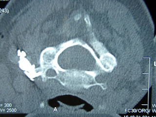
This patient had neck pain on the left side irradiating to the scapular region after having been treated with a violent cervical rotation manipulation for a torticollis three years earlier. Her MRI demonstrated some insignificant disc bulging at the C3/4 level. The radiologist doing the MRI proposed a CT guided transforaminal injection of steroids which he subesequently performed as illustrated in the next two images.
 The needle has been introduced from behind on the left side towards the transverse process of C3. The root hole opens laterally-downwards. An appropriate needle pass would have been 90 degrees to the actual needle via an anterior approach and going slightly cranially, sliding up along the posterior/inferior aspect of the root hole, caudal to the ganglion and posterior to the vertebral artery. The needle tip shold not pass beyond the mid-width of the articular collumn as seen in a perfect a-p projection.
  Injection of contrast fails to reach the target; there is no penetration into the spinal canal and the nerve root is not being visualized. Contrast spreads backwards along the needle through the regions of the anterior, median and posterior scalene muscles and the splenius muscle of the head.
 Nedless to say, there was no therapeutic effect of the steroids. The patient was later diagnosed in our centre with pain from the left C5/6 and C6/7 zygapophysial joints. She was treated with radiofrequency neurotomy of the medial branches responsible for the innervation of these joints and her pain has now stabilized, 6 months after treatment, at a very acceptable level of 1-2/10, down from 7-8/10 before therapy.

No comments:
Post a Comment